Compared to other classes of proteins, determining membrane protein structures remains a challenge in large part due to the difficulty in establishing experimental conditions that can preserve the correct conformation of the protein in isolation from its native environment (Figure 2.33). Many roles of linear motifs are associated with cell regulation, for instance in control of cell shape, subcellular localisation of individual proteins and regulated protein turnover. 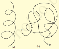 In Wikipedia, The Free Encyclopedia. (B) Structure of the tetrameric hemoglobin protein containing a total of four globin folds. 0000003082 00000 n
In nature, some proteins are formed from several polypeptides, also known as subunits, and the interaction of these subunits forms the quaternary structure. 4-6). The hexamer is an inactive form with long-term stability, which serves as a way to keep the highly reactive insulin protected, yet readily available. For the right-handed alpha helix, every helical turn has 3.6 amino acid residues (Figure 2.19). However, due to the proximity of, and electron withdrawing nature of the aromatic ring structure, the lone pair of electrons on the nitrogen are unavailable to accept a proton. In the -pleated sheet, the pleats are formed by hydrogen bonding between atoms on the backbone of the polypeptide chain. Figure 2.26 The TIM Barrel. The -OH groups of hydroxyproline are Figure 2.10 Formation of the Peptide Bond. Actually, the final beta-pleated sheet structure of silk is Cysteine is also a unique amino acid as this side chain is capable of undergoing a reversible oxidation-reduction (redox) reaction with other cysteine residues creating a covalent disulfide bond in the oxidized state (Figure 2.9). In some cases, hydrophobic clusters in disordered sequences provide the clues for identifying the regions that undergo coupled folding and binding (refer to biological roles). Understand the significance of antigen structure. Only Gly residues can be accommodated at the very tight junctions between the individual chains (above b). In, https://en.wikipedia.org/w/index.php?title=TIM_barrel&oldid=899459569, Wikipedia contributors. (A) -hairpin- structures are characterized by a sharp hairpin turn that does not disrupt the hydrogen bonding of the two -pleated sheet structures. The hydrophobic effect (Figure 2.37) is the phenomenon in which the hydrophobic chains of a protein collapse into the core of the protein (away from the hydrophilic environment). Intrinsically Unstructured Proteins (IUPs) occupy the extreme end of this spectrum of flexibility, whereas IDPs also include proteins of considerable local structure tendency or flexible multidomain assemblies. Figure 2.12 Cis and Trans Conformation of Amino Acid R-Groups. Figure 2.20 Left Handed Alpha Helix Structure. Each amino acid can be abbreviated using a three letter and a one letter code. 0000011701 00000 n
High concentrations of solutes, extremes of pH, mechanical forces, and the presence of chemical denaturants can contribute to protein denaturation, as well. About four protofibrils32 strands of -keratin in allcombine to form an intermediate filament. Enzymes are proteins that catalyze a reaction, and coenzymes are the non-protein molecules or elements that assist the process. Furthermore, the C-N bond within the amide structure is fixed in space and cannot rotate due to the pi-bond character. to normal upon relaxation of tension. Learn the fibroblast definition and understand its purpose. Disulfide bridges add additional stability to the 3-D structure and are often required for correct protein folding and function (Figure 2.14). On cooling, they spontaneously revert to the -helix conformation. Become a Study.com member to unlock this answer! The sheet of eight tetramers is then twisted into a lefthanded helix forming the final intermediate filament (E) An electron micrograph of the intermediate filament is shown in the upper lefthand corner. Intrinsic disorder is particularly enriched in proteins implicated in cell signaling, transcription and chromatin remodeling functions. (2016) PLosONE 11(3):e0151431. proteins. As seen in Table 2.1, seven of the amino acids contain R-groups with ionizable side chains and are commonly found in the active sites of enzymes. A very interesting factor about insulin is the three disulfide Learn about the catecholamines called epinephrine and norepinephrine that are released by the adrenal medulla when you are adapting to stress. The hexamer form of insulin is a way for the body to store insulin in a stable and inactive conformation so that it is available for release and reactivation in the monomer form. Registration peptides are cleaved and tropocollagen is formed by procollagen peptidase. Within each protein small regions of the protein may adopt specific, repeating folding patterns. Disordered regions are often found as flexible linkers or loops connecting domains. Integral membrane proteins are permanently attached to the membrane. The five most common types are: Here we will focus on the unique attributes of Collagen Type I. Collagen Type I has an unusual amino acid composition and sequence: Figure 2.30. (2019) Integumentary Levels of Organization. Thus, for simplicity sake, the 20 amino acids used for protein synthesis have both three letter and one letter code abbreviations (Table 2.1). The existence and kind of protein disorder is encoded in its amino acid sequence. In 1950, Karush wrote about Configurational Adaptability contradicting all the assumptions and research in the 19th century. Insulin starts out as a single polypeptide and loses some internal sequences during cellular processing that form two chains held together by disulfide linkages as shown in figure 2.14. In the hardest and toughest -keratins, such as those of rhinoceros horn, up to 18% of the residues are cysteines involved in disulfide bonds. 0000007250 00000 n
The polar, hydrophilic amino acids can be subdivided into three major classes, the polar uncharged-, the acidic-, and the basic- functional groups.
In Wikipedia, The Free Encyclopedia. (B) Structure of the tetrameric hemoglobin protein containing a total of four globin folds. 0000003082 00000 n
In nature, some proteins are formed from several polypeptides, also known as subunits, and the interaction of these subunits forms the quaternary structure. 4-6). The hexamer is an inactive form with long-term stability, which serves as a way to keep the highly reactive insulin protected, yet readily available. For the right-handed alpha helix, every helical turn has 3.6 amino acid residues (Figure 2.19). However, due to the proximity of, and electron withdrawing nature of the aromatic ring structure, the lone pair of electrons on the nitrogen are unavailable to accept a proton. In the -pleated sheet, the pleats are formed by hydrogen bonding between atoms on the backbone of the polypeptide chain. Figure 2.26 The TIM Barrel. The -OH groups of hydroxyproline are Figure 2.10 Formation of the Peptide Bond. Actually, the final beta-pleated sheet structure of silk is Cysteine is also a unique amino acid as this side chain is capable of undergoing a reversible oxidation-reduction (redox) reaction with other cysteine residues creating a covalent disulfide bond in the oxidized state (Figure 2.9). In some cases, hydrophobic clusters in disordered sequences provide the clues for identifying the regions that undergo coupled folding and binding (refer to biological roles). Understand the significance of antigen structure. Only Gly residues can be accommodated at the very tight junctions between the individual chains (above b). In, https://en.wikipedia.org/w/index.php?title=TIM_barrel&oldid=899459569, Wikipedia contributors. (A) -hairpin- structures are characterized by a sharp hairpin turn that does not disrupt the hydrogen bonding of the two -pleated sheet structures. The hydrophobic effect (Figure 2.37) is the phenomenon in which the hydrophobic chains of a protein collapse into the core of the protein (away from the hydrophilic environment). Intrinsically Unstructured Proteins (IUPs) occupy the extreme end of this spectrum of flexibility, whereas IDPs also include proteins of considerable local structure tendency or flexible multidomain assemblies. Figure 2.12 Cis and Trans Conformation of Amino Acid R-Groups. Figure 2.20 Left Handed Alpha Helix Structure. Each amino acid can be abbreviated using a three letter and a one letter code. 0000011701 00000 n
High concentrations of solutes, extremes of pH, mechanical forces, and the presence of chemical denaturants can contribute to protein denaturation, as well. About four protofibrils32 strands of -keratin in allcombine to form an intermediate filament. Enzymes are proteins that catalyze a reaction, and coenzymes are the non-protein molecules or elements that assist the process. Furthermore, the C-N bond within the amide structure is fixed in space and cannot rotate due to the pi-bond character. to normal upon relaxation of tension. Learn the fibroblast definition and understand its purpose. Disulfide bridges add additional stability to the 3-D structure and are often required for correct protein folding and function (Figure 2.14). On cooling, they spontaneously revert to the -helix conformation. Become a Study.com member to unlock this answer! The sheet of eight tetramers is then twisted into a lefthanded helix forming the final intermediate filament (E) An electron micrograph of the intermediate filament is shown in the upper lefthand corner. Intrinsic disorder is particularly enriched in proteins implicated in cell signaling, transcription and chromatin remodeling functions. (2016) PLosONE 11(3):e0151431. proteins. As seen in Table 2.1, seven of the amino acids contain R-groups with ionizable side chains and are commonly found in the active sites of enzymes. A very interesting factor about insulin is the three disulfide Learn about the catecholamines called epinephrine and norepinephrine that are released by the adrenal medulla when you are adapting to stress. The hexamer form of insulin is a way for the body to store insulin in a stable and inactive conformation so that it is available for release and reactivation in the monomer form. Registration peptides are cleaved and tropocollagen is formed by procollagen peptidase. Within each protein small regions of the protein may adopt specific, repeating folding patterns. Disordered regions are often found as flexible linkers or loops connecting domains. Integral membrane proteins are permanently attached to the membrane. The five most common types are: Here we will focus on the unique attributes of Collagen Type I. Collagen Type I has an unusual amino acid composition and sequence: Figure 2.30. (2019) Integumentary Levels of Organization. Thus, for simplicity sake, the 20 amino acids used for protein synthesis have both three letter and one letter code abbreviations (Table 2.1). The existence and kind of protein disorder is encoded in its amino acid sequence. In 1950, Karush wrote about Configurational Adaptability contradicting all the assumptions and research in the 19th century. Insulin starts out as a single polypeptide and loses some internal sequences during cellular processing that form two chains held together by disulfide linkages as shown in figure 2.14. In the hardest and toughest -keratins, such as those of rhinoceros horn, up to 18% of the residues are cysteines involved in disulfide bonds. 0000007250 00000 n
The polar, hydrophilic amino acids can be subdivided into three major classes, the polar uncharged-, the acidic-, and the basic- functional groups.
Secondary structure elements: -helices (red), -strands (blue arrows). Protein folding must be thermodynamically favorable within a cell in order for it to be a spontaneous reaction. Figure 2.35 Conformational flexibility in SUMO-1 protein (PDB:1a5r). Figure 2.38 Formation of a Water Clathrate. In some cases, IDPs can adopt a fixed three-dimensional structure after binding to other macromolecules. Figure 2.36 Protein Before and After Folding. In 1950, Karush wrote about Configurational Adaptability contradicting all the assumptions and research in the 19th century. The hydrophobic collapse introduces entropy back to the system via the breaking of the water cages which frees the ordered water molecules. Their structures, like their functions, vary greatly. A solution of a reducing agent, usually a compound containing a thiol or sulfhydryl group (SH), is then applied with heat. Absolute configuration. Fibrous Proteins are characterized by elongated protein structures. Retrieved 18:30, July 20, 2019, from https://en.wikipedia.org/w/index.php?title=Protein_folding&oldid=906604145, Wikipedia contributors. microfibril arrangement in which 5 triple helices are involved in a left-handed Glycine is needed because it is small and is the only amino acid The TIM barrel can also be thought of, then, as made up of 8 overlapping, right-handed -- super-secondary structures, as shown in the side view (B). 0000004720 00000 n is quite as effective as the human insulin. and most enzymes. However, as of 2011, 28 types of collagen have been identified, described, and divided into several groups according to the structure they form. The remaining oxygen atom is then used to hydroxylate an appropriate Pro residue in procollagen. Because myoglobin was the first protein whose structure was solved, the globin fold was thus the first protein fold discovered. The primary sequence of a protein is linked together using dehydration synthesis (loss of water) that combine the carboxylic acid of the upstream amino acid with the amine functional group of the downstream amino acid to form an amide linkage (Figure 2.10). 96 53 If the path is is in the clockwise direction, the chiral center is given the R-designation, whereas if the path is counterclockwise, it is given the S-designation. 0000106089 00000 n The R groups (the variant groups) of the polypeptide protrude out from the, Expanded Top View Linear Structure and Space-Filling Model, The alpha helix is more compact than the fully extended polypeptide chain with phi/psi angles of 180. This creates fixed physical locations of the R-groups within the growing peptide in either the cis or trans conformations. Figure modified from: Khara, J.S., et al. When left-handed helices do form, they are often critical for the correct protein folding, protein stability, or are directly involved in the formation of the active site. The hydrogen (2017) Acta Biomat 57:103-114 and Ryu, H., et al. Genes Overview, Types & Function | What are Genes? Within these structures, intramolecular interactions, especially hydrogen bonding between the backbone amine and carbonyl functional groups are critical to maintain 3-dimensional shape. startxref 1. a single transmembrane -helix (bitopic membrane protein) 2. a polytopic transmembrane -helical protein 3. a polytopic transmembrane -sheet protein. 300nm for a single collagen molecule. 0000004618 00000 n It does not denote the absolute stereo configuration of a molecule. Gly residues are shown in red. Retrieved 16:01, July 20, 2019, from https://en.wikipedia.org/w/index.php?title=Rossmann_fold&oldid=904468788, Wikipedia contributors. The multitude of hydrophobic groups interacting within the core of the globular folded protein contributes a significant amount to protein stability after folding, because of the vastly accumulated van der Waals forces (specifically London Dispersion forces). Amino This is the case with the TIM Barrel, a conserved protein fold consisting of eight -helices and eight parallel -strands that alternate along the peptide backbone. fibril is the same distance. Understand more about chromatin's role in DNA repair, chromosomes, and examples of chromatin in this lesson. Multiple tropocollagen molecules form collagen fibrils, via covalent cross-linking (aldol reaction) by lysyl oxidase which links hydroxylysine and lysine residues. Thus, histidine will slowly progress from an overall +2 charge at very low pH (fully protonated) to an overall -1 charge at very high pH (fully deprotonated). Three of these structures are then grouped further forming an inactive hexamer (Figure 2.28). Transport proteins move molecules and ions across the membrane. Hydrogen bonding between the backbone carbonyl and the backbone amine functional groups stabilized both the antiparallel (B left) and the parallel (B right) -pleated sheet structures. Thus disordered sequences cannot sufficiently bury a hydrophobic core to fold into stable globular proteins. This is still speculative, but recent findings from meteorites make this hypothesis much more plausible. TIM barrels are one of the most common protein folds. The process of folding often begins co-translationally, so that the N-terminus of the protein begins to fold while the C-terminal portion of the protein is still being synthesized by the ribosome; however, a protein molecule may fold spontaneously during or after biosynthesis. Createyouraccount. What are the primary, secondary, and tertiary structures of collagen? Nuclear magnetic resonance spectroscopy of proteins also demonstrated the presence of large flexible linkers and termini in many solved structural ensembles. Click Here for a Downloadable Version of the Amino Acid Chart. sequence of: where X is any amino acid and hypro is hydroxyproline. Vitamin C is required for, the hydroxylation of proline and lysine in collagen; scurvy is a deficiency disease characterized by general degeneration of connective tissue. What is an extracellular matrix? Manifestations of advanced scurvy include numerous small hemorrhages caused by fragile blood vessels, tooth loss, poor wound healing and the reopening of old wounds, bone pain and degeneration, and eventually heart failure. Based on their shape, function and location proteins can be characterized broadly as fibrous, globular, membrane, or disordered. Explore how cellular adaptation happens during different types of cell growth, including hypertrophy, hyperplasia, and neoplasia. Thus, the R- and S-designations do not always correspond with the D- and L- conformation. In the example above the two peptide chains that form the hormone insulin are depicted. It is the physical process by which a polypeptide folds into its characteristic and functional three-dimensional structure from random coil. Conversely, the torsion angle Psi () measures the rotation around the -carbon carbonyl carbon bond by evaluating the angle between the two neighboring nitrogen atoms when you are looking directly down the -carbon carbonyl carbon bond (Figure 2.16). Two of them hold the two chains together, and the third Structural proteins, e.g., actin and tubulin, which are globular and soluble as monomers, but polymerize to form long, stiff fibers. Molecular model studies There are two major categories of Collagen may be attached to cell membranes via several types of protein, including fibronectin, laminin, fibulin and integrin. aligned such that the maximum electrostatic and hydrophobic interactions occur Chaperones exist in all cellular compartments and interact with the polypeptide chain in order to allow the native three-dimensional conformation of the protein to form; however, chaperones themselves are not included in the final structure of the protein they are assisting in. Retrieved 03:42, July 20, 2019, from https://en.wikipedia.org/w/index.php?title=Collagen&oldid=906509954, Wikipedia contributors. Figure 2.16 Phi () and Psi () Torsion Angles. Collagen is present as tropocollagen containing triple helix. head-to-tail, with a 35nm gap between each, in a staggered bundle as shown. Synthesis of Collagen Type I. Polypeptide chains are synthesized in the endoplasmic reticulum and released into the lumen where they are hydroxylated and glycosylated. These publications solidified the central dogma of molecular biology in that the sequence determines the structure which, in turn, determines the function of proteins. which is equivalent to 67 nm; while the gap between staggered molecules in a Parathyroid glands are endocrine glands on the posterior surface of the thyroid gland. The chart of standard pKa values for the amino acids is shown in Table 2.1 and can be used to predict the ionization/charge status of amino acids and their resulting peptides/proteins. In Wikipedia, The Free Encyclopedia. Thus, proteins are always synthesized in a directional manner starting with the amine and ending with the carboxylic acid tail. One of the most intriguing features among members of this class of proteins is although they all exhibit the same tertiary fold there is very little sequence similarity between them. Two types of alpha chains are formed during translation on ribosomes along the rough endoplasmic reticulum (RER): alpha-1 and alpha-2 chains. Retrieved 19:52, July 20, 2019, from https://en.wikipedia.org/w/index.php?title=Intrinsically_disordered_proteins&oldid=905782287, Medicinal Chemistry and Pharmacology Concentration. in new window. 0000002012 00000 n Fascia in Anatomy Overview & Function | What Is Fascia? (4)In -keratins, the cross-links stabilizing quaternary structure are disulfide bonds. In the lower diagram, any amino acid, X is positioned upstream of a proline residue. In cellular systems, disulfide bond formation/disruption is an enzyme-mediated reaction and can be utilized as a mechanism to control the activity of protein.
Learn the definition of the basal lamina and understand its main functions. The hair to be waved or curled is first bent around a form of appropriate shape. All the R-groups extend outward and away from the helix axis. Not surprisingly, -keratin is rich in the hydrophobic residues Ala, Val, Leu, Ile, Met, and Phe. of the insulin proteins. Depending upon the degree of mineralization, collagen tissues may be rigid (bone), compliant (tendon), or have a gradient from rigid to compliant (cartilage). The resulting three-dimensional structure is determined by the amino acid sequence or primary structure (Anfinsens dogma). utilization of glucose. Consider how the shape of this molecule might relate to its ability to In the hydrolysis reaction, water is added across the amide bond incorporating the -OH group with the carbonyl carbon and reforming the carboxylic acid. The helical conformation of each chain is dependent on the fact that every thirdresidue is a Gly, and that the sequence is rich in Pro. 0000001356 00000 n The overall structure is stabilized by extensive hydrogen bonding between all peptide linkages in the polypeptides of each sheet and by the optimization of van der Waals interactions between sheets. In, https://en.wikipedia.org/w/index.php?title=Globular_protein&oldid=901360467, Wikipedia contributors.
Examples of folding catalysts are protein disulfide isomerases and peptidyl-prolyl isomerases that may be involved in formation of disulfide bonds or interconversion between, Chaperones are shown to be critical in the process of protein folding, A fully denatured protein lacks both tertiary and secondary structure, however, the primary protein sequence remains intact and the protein exists as a random coil (Figure 2.39). Figure 2.14 Disulfide Bonds. Proteins will have limitations on their folding abilities by the restricted bending angles or conformations that are possible, as described by the Ramachandran plot. While the latter are rigid and contain only one set of Ramachandran angles, IDPs involve multiple sets of angles. helix (there are three segments). In Wikipedia, The Free Encyclopedia. Proteins can also act as structural scaffolding within the cell, helping to maintain cellular shape. In the 1930s -1950s, the first protein structures were solved by protein crystallography. Methionine, one of the sulfur-containing amino acids is usually classified under the nonpolar, hydrophobic amino acids as the terminal methyl group creates a thioether functional group which generally cannot form a permanent dipole within the molecule and retains low solubility. Intrinsically disordered proteins can retain their conformational freedom even when they bind specifically to other proteins. This is due to the Phi () and Psi () torsion angles required to obtain the left-handed alpha helical structure. It is measured as the angle between the two carbonyl carbon atoms adjacent to the bond, shown in the lower panel. The amino acids that play a significant role in the binding specificity of the active site are usually not adjacent to each other in the primary structure, but form the active site as a result of folding in creating the tertiary structure, as you will see later in the chapter. 2. Collagens are found in tendons and other connective ligaments. repeated sequence Gly-Pro-Pro in each of the 3 alpha-chains, subjected to an Two of these structures include twisted sheets or saddles as well as beta barrels (Figure 2.24), Figure 2.24 Common Beta Strand Structural Motifs. Due to its tightly wound structure, it can function as one of the strongest biological materials and has various uses in mammals, from predatory claws to hair for warmth. helical shape? The term globin can refer more specifically to proteins including the globin fold. Figure 2.11 Amide Resonance Structure. Linear motifs are short disordered segments of proteins that mediate functional interactions with other proteins or other biomolecules (RNA, DNA, sugars etc.). These sequences revealed that there are two distinct but homologous keratin families which were named as Type I keratin and Type II keratins.